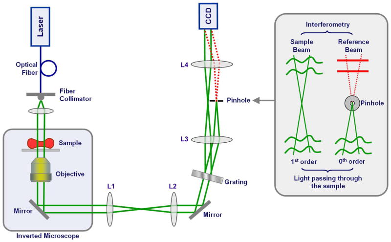
Figure 2.
DPM experimental setup. Collimated light from a 514 nm Argon laser is coupled to an inverted microscope (Olympus IX71) equipped with a high numerical aperture 40x objective. An amplitude grating generates multiple diffraction orders containing full spatial information about the sample image. The 0th order beam is low-pass filtered using a pinhole placed at the Fourier plane so that it becomes a plane wave after passing through lens L4. A common-path Mach-Zender interferometer is thus created with the 0th order as the reference beam and the 1st order as the sample beam (inset). Compared to conventional Mach-Zender interferometers, the two beams propagate through the same optical components, which essentially eliminates the phase noise without the need for active stabilization. Figure adapted from [28].

