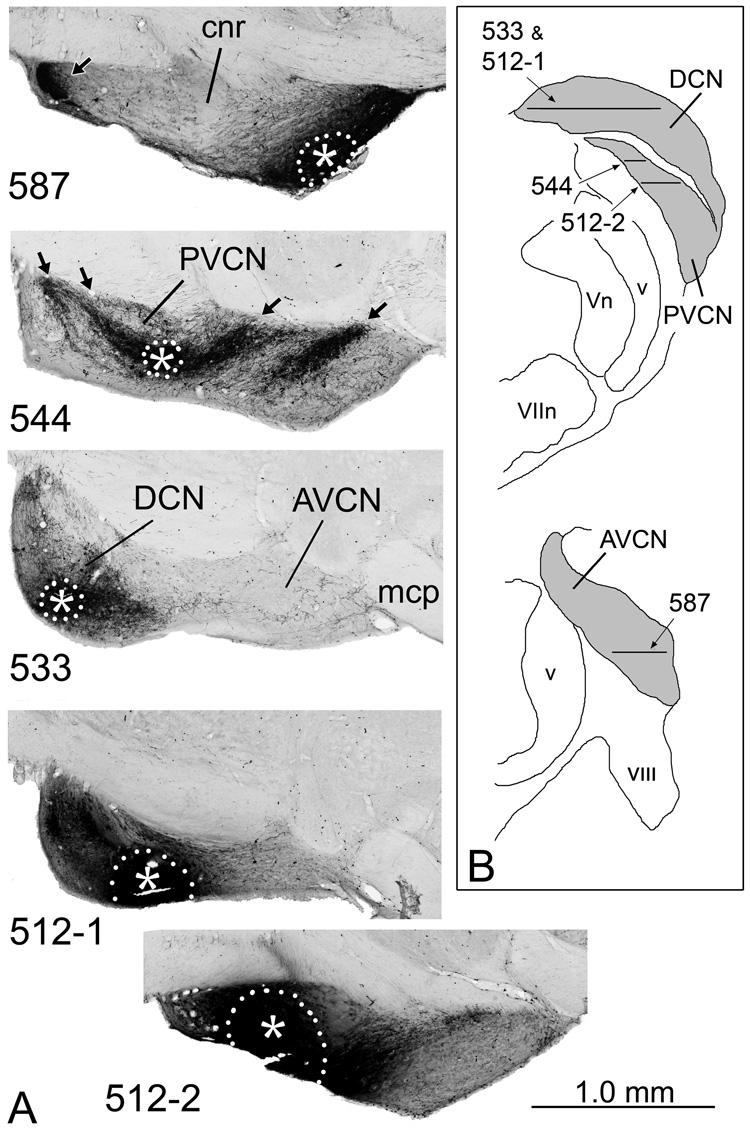
Figure 2.
A. BDA injection sites in the cochlear nucleus in four representative cases. The injection site in 587 was confined to the AVCN; that in 544, to the PVCN; and that in 533, to the DCN. In 512, distinct injection sites were located in both the DCN (512-1) and PVCN (512-2). The approximate dorsal-to-ventral position of each section is indicated on the drawings in panel B. The injection sites, which appear solid black because of accumulation of the BDA reaction product, are outlined by white dots with the approximate center indicated by asterisks. Terminal label in other subdivisions is evident (arrows) because the tracer is taken up by interneurons and auditory nerve fibers and transported to other parts of the complex. In this and subsequent figures, illustrations of sections through case 587, in which the injection site was on the left side, are rotated about the midline for easier comparisons to the other cases, in which the injection sites were on the right. In these horizontal sections, rostral is toward the right and medial is toward the top. The figure was assembled in Adobe Photoshop. Brightness and contrast were adjusted globally and background elements outside the brainstem were erased. B. Drawings of transverse sections through the caudal (top) and rostral (bottom) cochlear nuclear complex (indicated by gray fill) to illustrate the positions of the sections shown in panel A. Lateral is toward the left; dorsal is toward the top.

