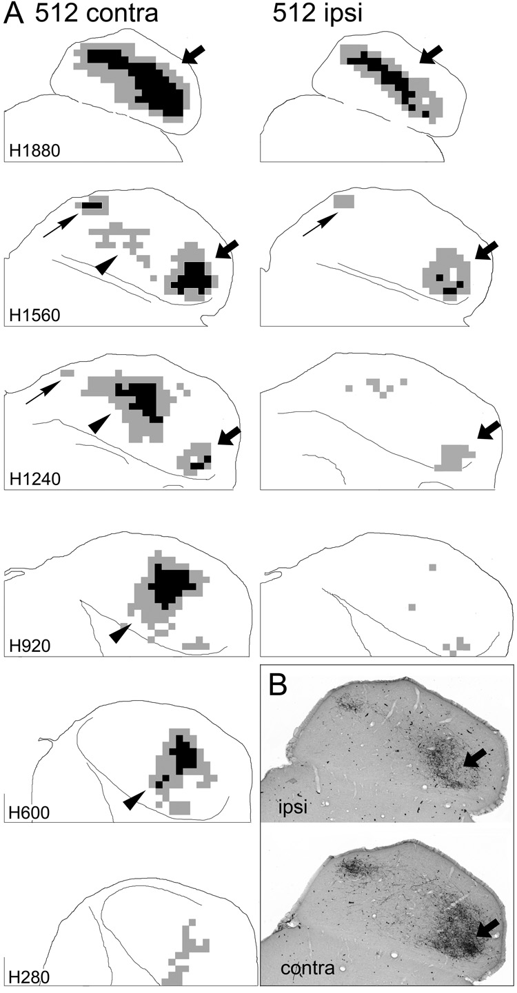
Figure 5.
Representation of terminal label in the ipsilateral (ipsi) and contralateral (contra) inferior colliculi in one case in which distinct injection sites were located in both the DCN and PVCN. A. Sections from the two sides are arranged as in Figure 3 and Figure 4 except that the most ventral levels are not shown for the ipsilateral IC. (Levels more ventral than those shown contained very little ipsilateral labeling.) The sections through the ipsilateral IC were rotated about the midline so that the results from the two sides are more easily compared. Arrows indicate patches of labeled elements as in Figure 3 and Figure 4 and are referenced where appropriate in the text. B. Digital images at one horizontal level (comparable to atlas level H1720) through the IC in the same case shown in panel A. The arrows indicate a comparable area on the two sides that exhibits heavy terminal labeling contralaterally but not ipsilaterally.

