Figure 2.
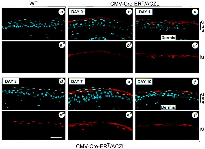
Pattern of Cre-ERT expression in the tail epidermis of double transgenic mice. Immunohistochemistry with anti-Cre antibody was performed on sections (10 μm-thick) of tail biopsies of 6- to 8-week-old wild-type (WT; a, a′) and CMV-Cre-ERT/ACZL double heterozygous transgenic mice (b–f, b′–f′). Double transgenic mice were injected for 5 consecutive days (days 0–4) with tamoxifen (1 mg/day). Sections were stained with DAPI and anti-Cre antibody. DAY 0 (b, b′), before the first tamoxifen injection; DAY 1 (c, c′) and DAY 3 (d, d′), after 1 and 3 days of tamoxifen treatment, respectively; DAY 7 (e, e′) and DAY 10 (f, f′), 3 and 6 days after the last tamoxifen injection, respectively. The cyan color corresponds to the DAPI staining (a–f), and the red color corresponds to the staining of Cre-ERT (a–f, a′–f′). B, S, and G, basal, spinous, and granular layers, respectively (see Fig. 1). (Bar = 25 μm.)

