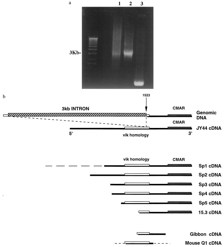
Figure 4.
Genomic organization of CMAR. (a) PCR from (as shown in Fig. 1) genomic DNA, (as shown in Fig. 2) cosmid DNA, and (as shown in Fig. 3) cDNA by using primers JY44F5P3 and JY44R1. Lambda one kilobase markers are shown on the left. (b) Human genomic DNA showing position of intron compared with human cDNAs, JY44, Sp1, Sp2, Sp3, Sp4, Sp5, and 15.3, gibbon cDNA, and mouse cDNA Q1. The broken line at the 5′ end of Sp1 indicates rearranged sequence. The dotted lines in mouse Q1 cDNA indicate no homology with other sequences. The vik homology region and the originally reported CMAR region are shown.

