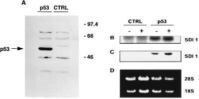
Figure 7.
pTpT up-regulates the expression of SDI1 mRNA through p53. Transfections of p53-null H1299 cells were carried out as described in Materials and Methods. (A) One day after transfection, cells were collected for Western blot analysis using 20 μg total protein as described (23). (B–D) Duplicate cultures of H1299 cells were transfected with the p53 expression vector (designated p53) or control vector (Ctrl) were given either diluent (DMEM) (designated −) or 100 μM pTpT (designated +). Northern blot analysis was performed with the SDI1 cDNA probe. The autoradiograph was scanned, and the brightness and contrast were adjusted to maximize differences in autoradiographic signals (B and C). (D) The ethidium bromide-stained gel is shown to demonstrate relative RNA loading. Densitometric analysis of the hybridization signals showed a >2-fold increase in the level of SDI1 mRNA by pTpT in the p53-transfected cells after correction for slight underloading of the +p53/+pTpT vs. +p53/−pTpT (D vs. C) lanes.

