Figure 4.
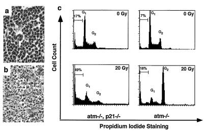
atm/p21 double-null tumors exhibit increased apoptosis. (a and b) Hematoxylin and eosin staining of tumor section. ×40 magnification. (a) An atm thymic lymphoma shows uniform T lymphoblastic cells with no detectable signs of apoptosis. (b) An atm/p21 double-null thymic lymphoma shows extensive apoptosis. (c) DNA damage-induced defective G2/M checkpoint control in atm/p21 double-null thymomas leads to increased apoptosis in thymic tumor cells. Tumor cell lines were irradiated at 0 and 20 Gy. Cells were collected at 24 hr, fixed and stained with propidium iodide, and analyzed for DNA content and apoptotic cells. G1 and G2 peaks are indicated as well as percentage of apoptotic cells. Similar observations were seen at 5 and 10 Gy of irradiation and from independent cell lines (not shown).

