Figure 2.
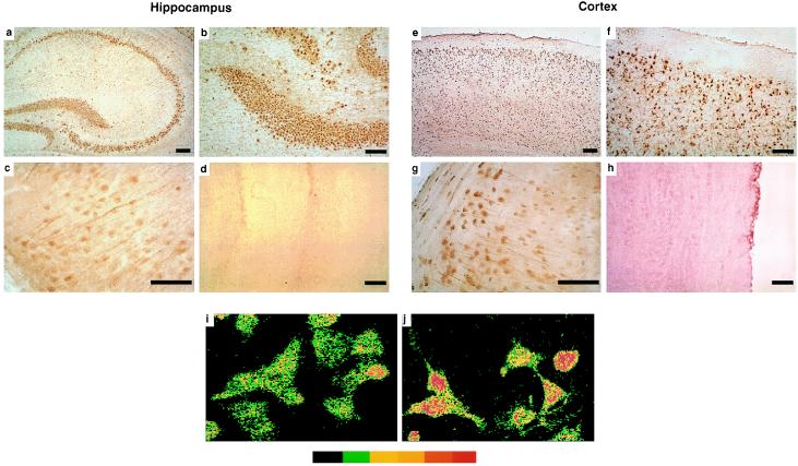
Neuronal expression of JNK immunoreactivity in brain and cell culture. (a–h) Cryosections (10 μm) of hippocampus (a–d) and neocortex (e–h) of unmanipulated mice were incubated with either JNK rabbit polyclonal antibody (Santa Cruz Biotechnology) (a–c, e–g, and i–j) or JNK antibody preadsorbed with recombinant JNK protein (d and h). Primary antibody binding was detected with species-matched secondary antibodies and the immunoperoxidase reaction using ABC reagents from Vector Laboratories and diaminobenzidine as the chromagen (brown reaction product). Note the widespread immunostaining of neuronal cell populations in the hippocampus and neocortex. Some neurons showed stronger nuclear staining (g). No cellular immunolabeling was detected with preadsorbed JNK antibody (d and h). (i–j) Neuro-2a cells in culture either were left untreated (i) or irradiated for 10 min with UV-C (j). Pseudocolor indicates increasing levels of immunofluorescence (from left to right) as determined by confocal microscopy. UV-C irradiation resulted in increased nuclear immunostaining for JNK. This was paralleled in replicate cultures by an increase in JNK enzymatic activity (Fig. 1c, Upper). (Bar: 250 μm.)

