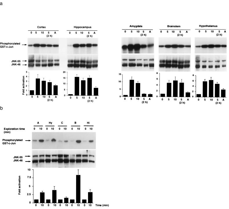
Figure 3.
Noninvasive environmental stimulation induces prominent JNK activation in mouse brain. (a) Physical restraint. Mice were unmanipulated (0) or restrained for 5 min (5), 10 min (10), or for 5 min followed by a 2-hr recovery period [5 (2h)]. Additional mice (n = 3) were killed by metofane anesthesia (A). (Top) Representative autoradiographs revealing JNK activities in five of the brain regions analyzed. (Middle) Western blot analysis of JNK protein levels in same tissue lysates. (Bottom) For each brain region, the average baseline level of JNK activity found in unmanipulated mice (n = 3) was arbitrarily defined as 1.0. Fold activation indicates average increases in JNK activity over baseline levels determined by PhosphorImager quantitations of signals obtained in manipulated mice (n = 3/group) in the same kinase assay (error bars = SD). Similar results were obtained in two additional independent kinase assays. (b) Exploration of a novel environment. Control mice were set down into the new cage for only 2–3 sec and returned immediately to their home cage for a 10-min observation period (0), whereas experimental mice were allowed to explore the new environment for 10 min (10). Representative autoradiographs revealing JNK activities (Top) and JNK protein levels (Middle), respectively. (Bottom) For each brain region, the mean level of JNK activity found in control mice (n = 4) was arbitrarily defined as 1.0. Fold activation indicates average increases in JNK activity over control levels determined by PhosphorImager quantitations of signals obtained in experimental mice (n = 4/group) (error bars = SD). Similar results were obtained in an additional independent kinase assay.

