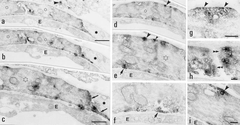
Figure 4.
Electron micrographs of arterioles. (a–c) A Y1-R+ (open stars) and a Y1-R-negative (stars) SMC in two adjacent sections. The Y1-R-LI is distributed in the cytoplasm and more intensely at lateral portions (arrows) of the SMC. The scattered labeling (arrowhead) in the cell peripheral to the SMC is unspecific. (c) Higher magnification from b. (d–i) Several intensely Y1-R-Ir sites are seen along the plasmalemma facing the perivascular space (arrowheads), the endothelium (arrows), or laterally toward another SMC (double arrowheads). Note close association with vesicles in many cases. E, endothelium. [Bars = 1 μm (a, b, and d), 500 nm (c and e), 250 nm (f and i; g and h).]

