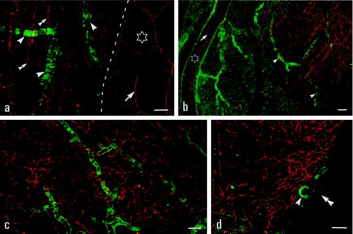
Figure 5.
Confocal micrographs showing Y1-R (green) and NPY (red) -LIs. (a) NPY+ nerve fibers (arrows) but no Y1-R-LI are seen around the inferior anterior cerebral artery (stars). Strong Y1-R-LI around some small arterioles directly derived from the basilar artery, but no NPY+ nerve fibers are seen along these arterioles. Double arrowheads pointing to central NPY+ nerve fibers. (b) Strongly Y1-R-Ir arterioles lacking NPY+ nerve fibers (arrowheads). NPY+ fibers are seen around a first branch (stars) of the middle cerebral artery. (c) A dense network of central NPY+ nerve fibers intermingles with strong Y1-R+ arterioles (basal cortex). (d) A Y1-R+ superficial arteriole (arrowheads) adjacent to NPY+ nerve fibers in the basal hypothalamus. The pia mater (double arrowheads) is not labeled. [Bars = 25 μm (a, b), 50 μm (c), and 100 μm (d).]

