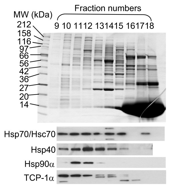
Figure 5.
Analysis of rabbit reticulocyte lysate by size separation and western blotting. Top, SDS-PAGE analysis of the peak fractions (9 to 18) from the size-exclusion separation of a crude rabbit reticulocyte lysate. The molecular weight standards for the SDS-PAGE are indicated on the left. Based on the molecular weight standards for the size exclusion column, fractions 9–11 are >700 kDa; fractions 12–13 are 600–200 kDa; fractions 14–20 are 200–10 kDa. Bottom, western blot analyses of these fractions (9–18) using antibodies against Hsp70/Hsc70, Hsp40, Hsp90a and TCP-1a.

