
Figure 2. Therapeutic Implications of Cardiac Progenitor Cells.
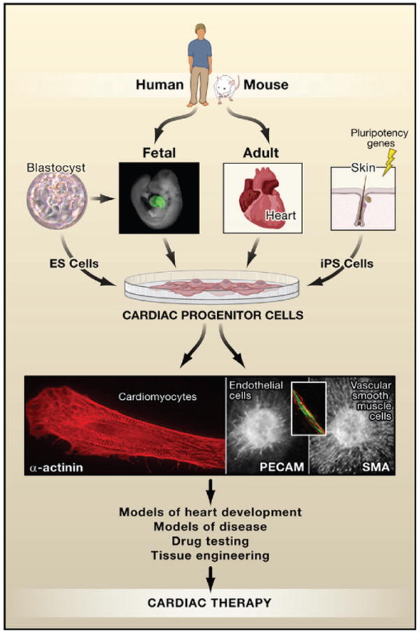
Progenitor cells have been described in fetal and adult heart in multiple species including humans. They also form as an intermediate in the differentiation of embryonic stem (ES) cells giving rise to cardiomyocytes, smooth muscle cells, and endothelial cells. Cardiac progenitors may be uni-, bi-, or tri- potent, depending on their molecular signatures although this relationship is still under investigation. Cardiomyocytes are morphologically readily identifiable by α-actinin staining (red, left panel) of sarcomeric structures, endothelial cells by expression of surface markers such as PE-CAM (green, middle panel and inset), and vascular smooth muscle cells that surround blood vessels by smooth muscle actin (sma; red, right panel). Cells expressing Nkx2.5 in the fetal mouse heart are indicated by green fluorescent protein (GFP) in a transgenic Nkx2.5-GFP mouse. Photos courtesy of C. Mummery (blastocyst; cardiomyocyte); F. Lebrin, D. Ward, and L. Tertoolen (endothelial cell and vascular smooth muscle cell); Sean Wu (fetal heart).