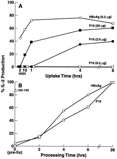
Figure 1.
Enhanced uptake of HBcAg particles vs. the P16 monomer by splenic APC. (A) Antigen uptake. The HBcAg and P16 antigens at the indicated concentrations were “pulsed” on unprimed B10.S (H-2s) mouse spleen cells as the source of APC for various lengths of time (2 min to 6 h) or 16 h for the control culture. After the antigen-pulse phase, splenic APC (3 × 10⁵) were vigorously washed (3×) and cultured with the IAs-restricted, HBcAg-specific T cell hybridoma 3E3–5G4 (1.2 × 10⁴) for 24 h, and culture supernatants (SN) were harvested. IL-2 production is expressed as a percentage of IL-2 induced by APC pulsed with HBcAg or P16 for 16 h. (B) Antigen processing. Splenic APC were loaded with HBcAg (0.1 μg/ml) peptide 120–140 (0.2 μg/ml) or P16 (50 μg/ml) (a greater concentration of P16 was necessary to compensate for the enhanced uptake of HBcAg) for 15 min, and washed. The splenic APC were either prefixed with glutaraldehyde (0.025% × 30 s) before antigen loading or at 2, 4, 6, or 26 h after antigen loading. Fixation inactivates the cellular metabolism required for antigen processing. The splenic APC fixed at various time points were then cultured with the 3E3–5G4 T hybridoma, and IL-2 production relative to the 26-h processing time was determined. These experiments were performed at least three times, and the results are representative with minor differences attributable to the use of different T cell hybridomas.

