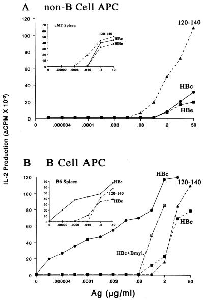
Figure 2.
B cells preferentially present the HBcAg to T cell hybridomas. (A) Spleen cells from unprimed B10(H-2b) mice were depleted of T cells and B cells and irradiated (2500 R) to inactivate any residual B cell APC function (11). Non-B cell APC (2 × 10⁵) were cultured with the IAb-restricted, HBcAg-specific T cell hybridoma 4E4–2B9 (1.2 × 10⁴) and media or the indicated concentrations of HBc, HBe, or the peptidic T cell site, amino acids 120–140. After 16 h, APC–T hybridoma culture SN was harvested and IL-2 was measured. (B) T cell-depleted spleen cells from unprimed B10 mice were enriched for B cells by removal of plastic adherent cells and passage of the nonadherent cells over a G-10 Sepherose column. A B cell myeloma cell line (HB99) was also used as a source of APC for HBcAg. B cell-enriched APC (2 × 10⁵) were cultured with the 4E4–2B9 T cell hybridoma and antigens as described above. The Insets depict identical experiments except that the source of APC was either spleen cells from B cell knockout μMT mice (A) or spleen cells from wild-type (+/+) B6 mice (B). The experiments using purified APC populations were performed with a number of different IAs and IAb-restricted HBcAg-specific T hybridoma cells on at least six occasions, and the results are representative.

