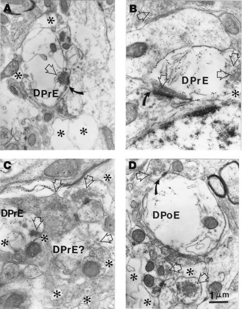
Figure 4.
Electron micrographs demonstrating degenerating pre- and postsynaptic elements (DPrE and DPoE, respectively) in an Atm knockout specimen, recognized by swelling, loss of cytoplasmic texture, and membrane-like remains in profiles participating in synaptic junctions recognized by their synaptic clefts (curved arrows) and, in the case of presynaptic elements (A–C), by some remaining synaptic vesicles (hollow arrows, used in all panels to highlight clusters of synaptic vesicles in both normal and degenerating profiles). Degenerating postsynaptic elements can be recognized by their associated synaptic clefts (curved arrow in B) to which a presynaptic profile containing synaptic vesicles (hollow arrow) is apposed. In addition to the pre- and postsynaptic degenerating profiles, all panels display an irregular, patchy loss of texture and disruption of the neuropil, which is scattered both around the degenerating profiles as well as around the nearby, otherwise apparently normal neuropil. The most severely disrupted neuropil is entirely devoid of protoplasm and organelles, containing only membrane-like remains, and is highlighted with asterisks in all panels.

