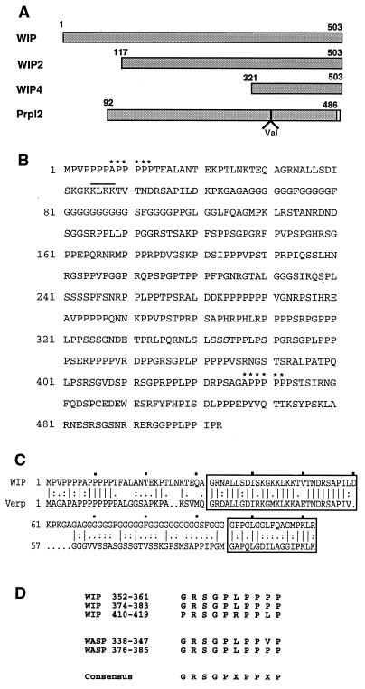
Figure 1.
WIP cDNAs, deduced amino acid sequence of WIP and its alignments. (A) Schematic representation of full-length WIP, WIP2, WIP4, and Prpl2 cDNAs. The open box in Prpl2 represents the seven amino acids that replace the C-terminal 17 amino acids in WIP. (B) Deduced amino acid sequence of WIP. The two APPPPP motifs implicated in profilin binding are denoted by ∗. A line is drawn over the KLKK motif implicated in acting binding. (C) Sequence alignment of the N-terminal regions of WIP and verprolin. The two verprolin homology regions are boxed. (D) Sequence alignment of GRSGPXPPXP motifs in WIP and WASP. Numbers refer to amino acid positions.

