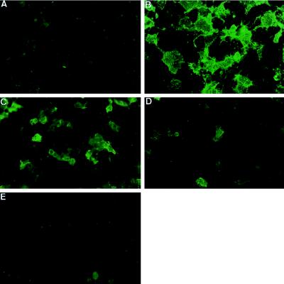
Figure 4.
Cell surface staining of transfected COS cells. IB4 lectin staining of cell surface of COS cells after: (A) mock transfection, (B) transfection with α1,3-galactosyltransferase cDNA, (C) transfection with α1,3-galactosyltransferase plus α-galactosidase cDNAs, (D) transfection with α1,3-galactosyltransferase plus α1,2-fucosyltransferase cDNAs, and (E) transfection with α1,3-galactosyltransferase plus α1,2-fucosyltransferase plus α-galactosidase cDNAs. Results are representative of at least 10 experiments.

