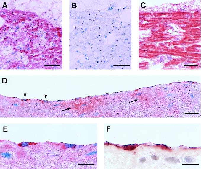
Figure 2.
Immunohistochemical demonstration of L-PGDS (β-trace) in the left atrium (A, B, D, E, and F) and right ventricle (C) of human heart. The tissue sections were immunostained with monoclonal antibodies against L-PGDS (A, C, D, and E) or von Willebrand factor (F), or with nonimmunized mouse IgG (B). (D) Arrowheads and arrows indicate the L-PGDS-immunoreactive endocardial cells and the immunoreactivity accumulated in the extracellular matrix, respectively. (Bars = 50 μm in A and B, 20 μm in C and D, and 10 μm in E and F.)

