Figure 3.
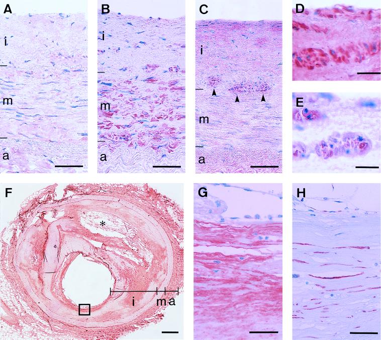
Immunohistochemical demonstration of L-PGDS (β-trace) in the left (A and B) and right (C, F, G, and H) coronary arteries and the right ventricle (D and E) of human heart. The tissue sections were immunostained with monoclonal antibodies against L-PGDS (A, C, D, F, and G) and alpha-smooth muscle actin (B, E, and H). (C) Arrowheads indicate the early phase of an arteriosclerotic plaque in the intima. (F) In the advanced atherosclerotic coronary artery, calcification (∗) and a lipid core including cholesterol clefts are observed. (G) A high-magnification view of a fibrous cap (squared in F). (Bars = 50 μm in A, B, C, G, and H; 20 μm in D and E; and 500 μm in F.) i, Intima; m, media; a, adventitia.

