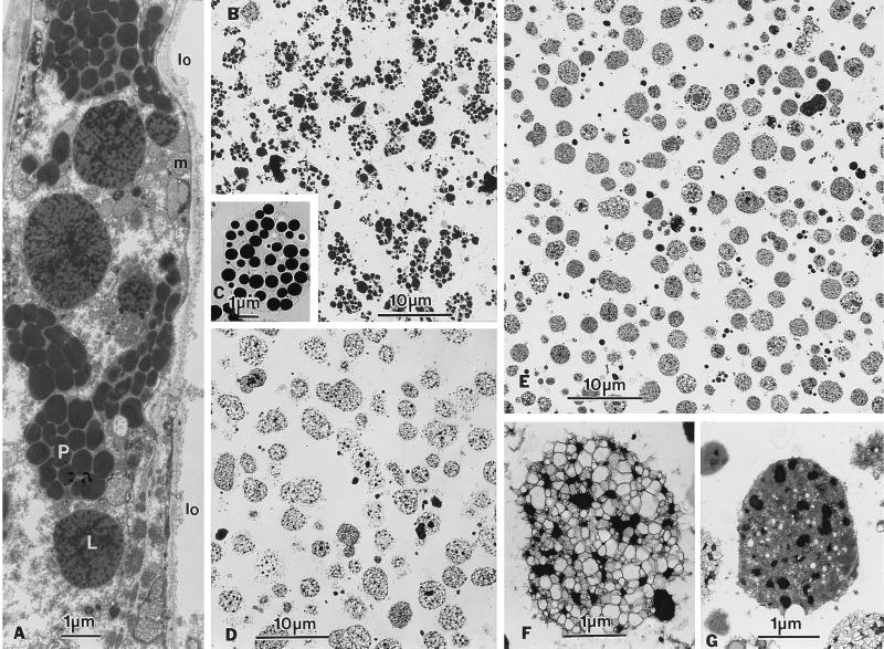
Figure 3.
Electron micrographs of a B. napus tapetum cell and isolated fractions of the plastids and lipid particles. (A) Portion of a tapetum cell, with its long axis facing the locule (lo). The cell contained abundant and conspicuous globuli-filled plastids (P) and lipid particles (L), as well as several mitochondria (m). (B) Fraction of isolated plastids, each of which was filled with globuli. (C) Enlarged isolated plastid, with a barely visible enclosing membrane. (D) Fraction of isolated lipid particles, aldehyde-fixed in a diluted osmotic solution. Most of the lipid particles were in a dilated form. (E) Fraction of isolated lipid particles, aldehyde-fixed in a higher osmotic solution. The lipid particles were in a dilated or dense form. In both D and E, the lipid particles in the dilated form were, in general, larger than those in the dense form. (F) Enlarged view of a lipid particle in the dilated form. Patches of osmiophilic materials, presumably neutral lipids, were scattered among vesicles. (G) Enlarged view of a lipid particle in the dense form.

