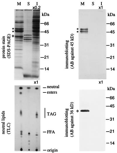
Figure 4.
SDS/PAGE and immunoblotting of proteins and TLC of neutral lipids of the two-organelle mixture (lane M), the ether-washed pollen surface materials (pollenkitt) (lane S), and the extract of the pollen interior after the ether wash (lane I) from B. napus. Proteins on the PAGE gel were stained with Coomassie blue (Upper Left) or treated for immunoblotting using antibodies against the 45-kDa polypeptide (Upper Right) or the 36-kDa polypeptide (Lower Right). Neutral lipids on the TLC plate were allowed to react with sulfuric acid (Lower Left). Positions of markers for protein molecular masses and standard lipids were shown on the right. Asterisks indicate the 48- and 45-kDa oleosins, and circles denote the plastid 36-kDa polypeptide.

