Figure 3.
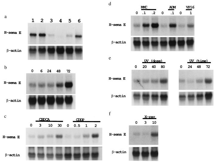
Induction of H-sema E mRNA in CDDP-sensitive cells. (a) Induction of H-sema E in CDDP-sensitive TYKnu cells by CDDP treatment. TYKnu cells were treated for 3 days with 0.0 (untreated control, lane 3), 0.1 (lane 4), 0.3 (lane 5), or 1.0 (lane 6) μg/ml CDDP. The constitutive expression of H-sema E by CDDP-resistant TYKnuR cells (lane 1) was not affected by the 3-day treatment with 1.0 μg/ml CDDP (lane 2). The relative intensity of the blots compared with lane 3 was 5.4 (lane 1), 6.3 (lane 2), 0.2 (lane 4), 1.5 (lane 5), and 4.3 (lane 6). (b) Time-dependent induction of H-sema E in CDDP-sensitive Lu65 cells. Lu65 cells were untreated or treated with 2.0 μg/ml CDDP for 6 to 72 hr (hereafter, lanes marked “0” represent untreated controls). The relative intensity of the blots compared with the untreated control was 1.1 (6 hr), 1.0 (24 hr), 2.0 (48 hr), and 3.7 (72 hr). (c) Dose-dependent induction of H-sema E in CDDP-sensitive Lu65 cells by the platinum-containing compounds, CBDCA and CDDP. H-sema E was induced in a dose-dependent manner by 3-day treatment with 3–30 μg/ml CBDCA or 0.5–2.0 μg/ml CDDP. The relative intensity of the blots compared with the untreated controls was 1.8 (3 μg/ml CBDCA), 1.8 (10 μg/ml CBDCA), 3.4 (30 μg/ml CBDCA), 1.2 (0.5 μg/ml CDDP), 2.9 (1.0 μg/ml CDDP), and 5.7 (2.0 μg/ml CDDP). (d) Induction of H-sema E in CDDP-sensitive Lu65 cells by non-platinum-containing anti-cancer compounds. H-sema E was induced by 3-day treatment with 0.1 and 0.2 μg/ml MMC, 0.1 μg/ml ADM, and 1.0 μg/ml VP-16. The relative intensity of the blots compared with the untreated controls was 2.2 (0.1 μg/ml MMC), 2.7 (0.2 μg/ml MMC), 2.9 (0.1 μg/ml ADM), and 3.1 (1.0 μg/ml VP-16). (e) Time- and dose-dependent induction of H-sema E in CDDP-sensitive Lu65 cells by UV irradiation. Total RNA was extracted from Lu65 cells 3 days after UV irradiation at 20, 40, or 80 J/m2 (Left), or 24, 48, or 72 hr after UV irradiation at 80 J/m2 (Right). The relative intensity of the blots compared with the untreated controls was 1.1 (20 J/m2), 1.5 (40 J/m2), 2.5 (80 J/m2), 0.6 (24 hr), 2.0 (48 hr), and 3.2 (72 hr). (f) Induction of H-sema E in CDDP-sensitive Lu65 cells by x-ray irradiation. Total RNA was extracted from Lu65 cells 3 days after irradiation at a dose of 3 or 10 Gy. The relative intensity of the blots compared with the untreated control was 0.7 (3 Gy) and 3.9 (10 Gy).

