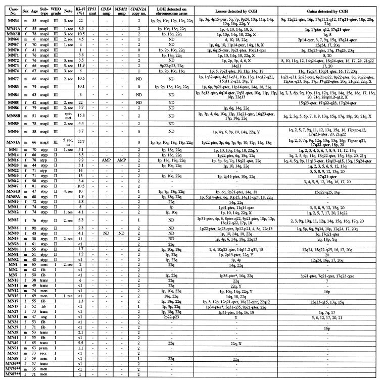
Figure 1.
Summary of selected clinical data, histopathological characteristics, and genomic alterations identified by CGH and other molecular genetic techniques in all meningioma groups investigated. m, Male; f, female; anapl, anaplastic; atyp, atypical; men, meningothelial; fib, fibrous; trans, transitional; ang, angiomatous; psam, psammomatous; secr, secretory; rec, recurrence; spin met, spinal metastasis; irr, irradiated prior to operation; mut, mutation; amp, amplification; ND, no data. High-level amplifications are given in boldface type in the section “gains detected by CGH.” ∗, Losses of 1p34–p36 determined by CGH were only included if confirmed by LOH data; ∗∗, meningiomas with brain invasion but no other signs of anaplasia.

