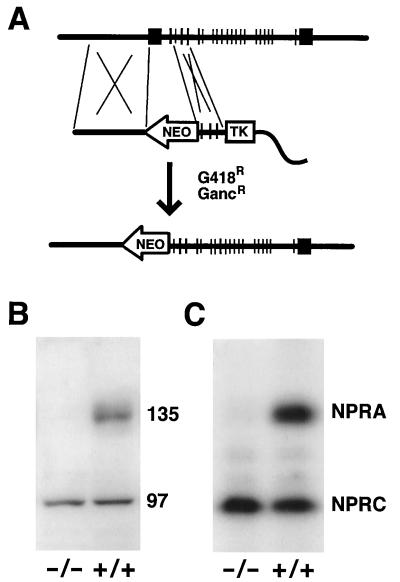
Figure 1.
Targeted disruption of the Npr1 gene. (A) The top line shows the structure of the Npr1 gene containing 22 exons spanning approximately 18 kb. The targeting construct (middle line) was designed to replace exon 1 through intron 1 with a neomycin resistance gene (NEO). TK indicates the Herpes simplex thymidine kinase gene, which with NEO allowed selection in a medium containing ganciclovir (Ganc) and G418. The cloning vector is represented by a wavy line. The bottom line shows the targeted locus. (B) A Western blot, using an antibody against the extracellular domain of rat NPRA, of the membrane fraction of kidney extracts prepared from Npr1 −/− (homozygous mutant) and +/+ (wild type) mice. The 135-kDa NPRA protein, present in the wild-type mice but absent in the mutants, is indicated, as is an unidentified cross-reacting protein of 97 kDa that is equally present in mice of both genotypes. (C) An autoradiogram showing the absence or presence of the 135-kDa NPRA after photoaffinity labeling with 4-azidobenzoyl [125I]-ANP in plasma membrane preparations of lung tissue from Npr1 −/− and wild-type (+/+) mice. The positions of the 135-kDa NPRA and 70-kDa natriuretic peptide receptor C radiolabeled bands are indicated.

