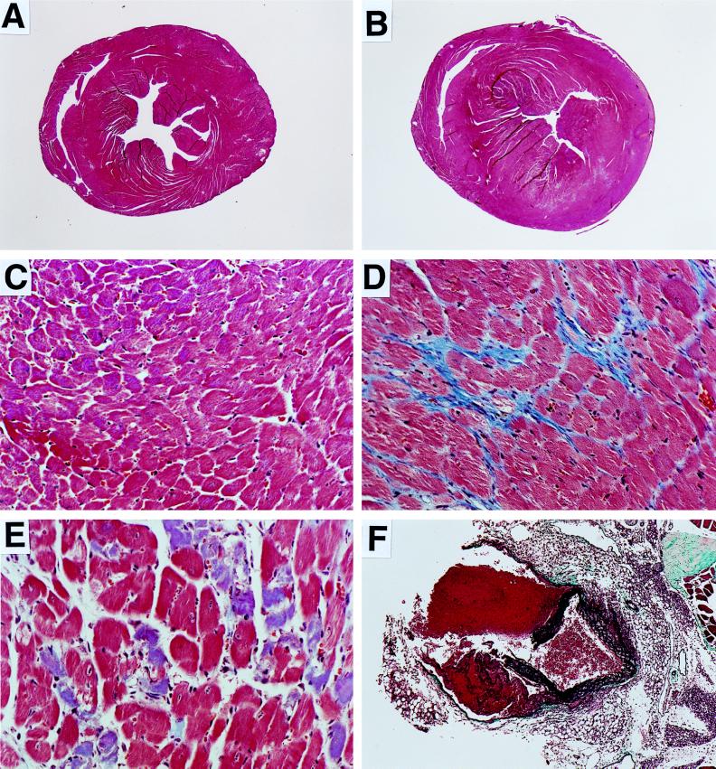
Figure 4.
Histological analysis of tissues from Npr1 −/− male mice and wild-type controls. (A–E) Heart tissues from euthanized animals stained with Masson’s trichrome. Hearts were fixed without diastole arrest and with no intracardiac pressure, consequently the in vivo dilatation of the Npr1 −/− hearts was not preserved. (A) ×9 and (C) ×175 magnification of transverse sections of the heart from a 3.5-month-old wild-type male. (B) ×9 and (D) ×175 magnification of sections from a 3.5-month-old Npr1 −/− male. Fibrotic regions stain blue. (E) ×175 magnification of a section of a 4-month-old Npr1 −/− male showing dying myocytes (blue). (F) ×45 magnification of the aortic dissection from a deceased 3.5-month-old Npr1 −/− male, stained with trichrome-elastin stain to show elastic and connective tissue. The platelet-fibrin strands in the clot in the lower left quadrant show that some bleeding had occurred before death of the mouse.

