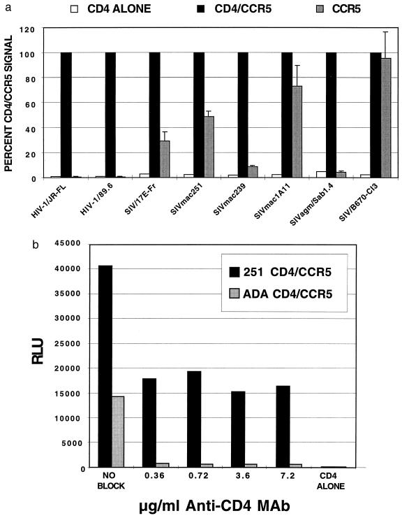
Figure 2.
(a) CD4-independent cell–cell fusion. Effector cells expressing the indicated viral envelope protein and T7 polymerase were mixed with target cells expressing luciferase under the control of the T7 promoter and CCR5 and CD4 as indicated. Values were normalized by expressing the signal as the percentage of signal obtained when CD4 and CCR5 were coexpressed (defined as 100% fusion) and represent the mean of three or four independent experiments with standard error bars shown. (b) Effects of an anti-CD4 MAb on cell–cell fusion. Cell–cell fusion was performed as in a. Effector cells expressing with the SIVmac251 or HIV-1 ADA env proteins were mixed with target cells expressing CD4 and CCR5 in the presence of MAb #19, a monoclonal antibody to CD4 that inhibits HIV-1 entry (16). The extent of cell–cell fusion was determined by measuring luciferase activity in relative light units (RLU) 5 h after cell mixing. The results are from a representative experiment.

