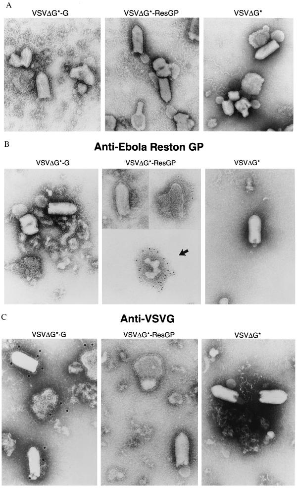
Figure 2.
Electron microscopy of recombinant VSV particles. VSVΔG* complemented with VSV G protein (VSVΔG*-G) or with ResGP (VSVΔG*-ResGP) or without complementation (VSVΔG*) was prepared and partially purified by centrifugation through 20% sucrose. Viruses were negatively stained (A) or labeled with a mouse mAb to ResGP (B) or VSV G protein (C) and anti-mouse IgG conjugated with colloidal gold, followed by negative staining.

