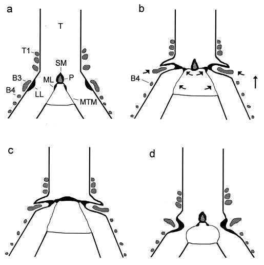
Figure 2.
(a) Configuration of the syrinx during quiet breathing in a frontal section (schematic). B3, B4, third and fourth bronchial ring; P, ossified pessulus; T, trachea; T1, first tracheal ring. (b) In the phonatory position the syrinx is moved rostrad and the bronchi are stretched (indicated by arrows), which, in concert with activity of the dorsal syringeal muscles, causes both labia to move mediad. LL is pushed into the lumen by B3, which presumably is rotated by dorsal muscle activity and also moved mediad by syringeal stretching. Movement of ML presumably is mediated by a thin, cartilaginous extension from the ventral end of B2 (not illustrated). (c) The dome-shaped phonatory position of the crow syrinx lacking an ossified pessulus. (d) The “classical model” of the phonatory position postulates constriction of the syrinx by the LL and vibrating MTM (redrawn after refs. 7 and 11).

