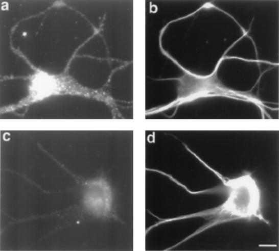
Figure 1.
Immunofluorescence micrographs of 4-day-old cultured cortical neurons double-labeled with trkC and a neuron specific tubulin monoclonal antibody. (a) Positive neuronal staining with trkC antibody and (b) corresponding staining with the tubulin antibody. (c) A negatively stained neuron with trkC antibody and (d) corresponding staining with neuronal specific tubulin antibody. (Bar = 10 μm.)

