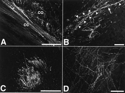
Figure 3.
Extensive axonal innervation of the host brain. The ES-cell-derived neurons generated a dense axonal network within the recipient brains. Abundant M6-positive axons were found at all levels in both gray and white matter. (A) Donor-derived axons in corpus callosum (cc) and deep layer cortex (co) of a 2-week-old recipient. (B) Axonal innervation of the hippocampal stratum oriens. The M6 immunofluorescence also depicts the perikaryon (arrow) and dendrites (arrowheads) of a large horizontal neuron in the upper stratum oriens. The morphology of this cell is very similar to the outline of Golgi-impregnated horizontal neurons in this area (20). (C) Abundant donor-derived axons in a striatal fiber tract of 2-week-old recipient brain. (D) ES-cell-derived axons in the thalamus of a newborn recipient transplanted at E17. (Bars = 50 μm.)

