Figure 5.
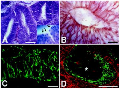
Generation of neuroepithelial formations. (A and B) Eight days after intrauterine transplantation, the donor cells have generated numerous neural tube-like structures within the host ventricle. Like the developing neural tube, these structures exhibit high mitotic activity at the luminal surface (A) (hematoxylin/eosin; arrows in Inset indicate mitotic figures) and strong expression of the intermediate filament nestin (B). (C and D) Neuroepithelial formation in the ventricle of a 2-week-old animal transplanted at E18. The formation contains abundant radially oriented nestin-positive processes (C). As in the early neuroepithelium, there is an inside-out gradient of differentiation with neuronal markers being expressed at the periphery of the formation (D) (green, tyrosine hydroxylase; red, M6; ∗, center of formation). [Bars = 100 μm (A and D) and 20 μm (B and C).]

