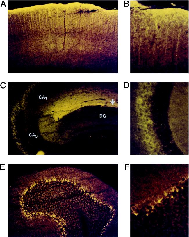
Figure 5.
Immunohistochemical analysis of BCNG-1 expression in the brain. Parasagittal sections of a mouse brain were stained with αq1 and αq2 antisera. The patterns of BCNG-1 expression detected with the two different antisera were identical, and in both cases the staining was entirely abolished by preadsorbing the sera with the GST-d5 fusion protein (not shown). (A, B) BCNG-1 immunoreactivity in the cerebral cortex. (C, D) BCNG-1 immunoreactivity in the hippocampus. In C, arrow shows position of hippocampal fissure; areas CA1, CA3, and dentate gyrus (DG) are labeled. D shows a detail of the stratum pyramidale of area CA3. (E, F) BCNG-1 immunoreactivity in the cerebellum. Magnification: ×100 (A,C,E); ×400 (B,D,F).

