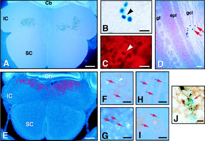
Figure 2.
(A–D) Transplant of postnatal X∷LacZ SVZ cells into the ventricle of E15 embryos. The recipient mouse was killed at P12 and the brain sectioned horizontally (animal 3-2, Table 1). X-Gal histochemistry produces a dark blue nuclear precipitate in X∷LacZ cells. In A and D, cell nuclei are counterstained with Hoechst 33258 and appear light blue under fluorescent illumination. (A) X-Gal+ cells in the IC. 490 cells were mapped in this 80-μm-thick section. (B) High-power view of the X-Gal+ cells in the inferior colliculus. (C) Example of X-Gal+ cell that was double-stained for the neuronal marker TuJ1 (arrowheads in B and C). (D) X-Gal+ cells in the granule layer of the olfactory bulb. (E–I) Transplant of postnatal NSE∷LacZ SVZ cells into the E15 ventricle. The recipient mouse was killed at P12 and the brain sectioned horizontally (animal 1-5, Table 1). An X-Gal perinuclear precipitate (typical examples indicated by red arrows) is produced in grafted cells which differentiate into neurons. All sections are counterstained with Hoechst 33258. (E) In this 80-μm-thick section, 790 X-Gal+ neurons were incorporated in the IC. Their mapped distribution is indicated by the red dots. (F) High-power view of E showing the graft-derived neurons in the IC. White arrowhead indicates nucleus corresponding to X-Gal positive cell. (G) Graft-derived neurons in the hypothalamus. (H and I) Graft-derived neurons in the olfactory bulb were found in the granule cell layer (H) and around glomeruli (star) (I). (J) Two GAD+/NSE∷LacZ+ neurons in the IC. The red arrow indicates the blue X-Gal deposit that became diffuse after the immunohistochemistry for GAD. The black arrows indicate a GAD+ cell body. Recipient killed at P1 (animal 9-1, Table 1). The localization of graft-derived cells in the IC varied from animal to animal (e.g., cells in A are more rostral, and in E more caudal). SC, superior colliculus; Cb, cerebellum; gl, glomerular layer; gcl, granule cell layer; epl, external plexiform layer. (Bars: A and E = 400 μm; D = 40 μm; B, C, F, G–I = 20 μm; J = 10 μm.)

