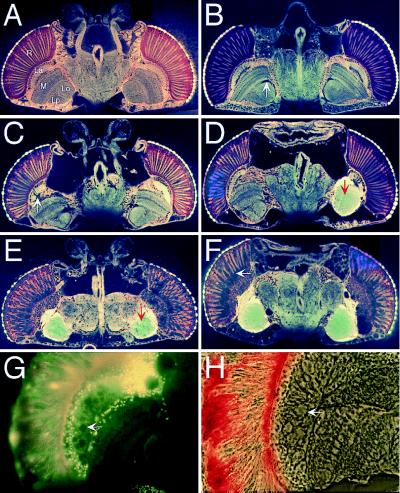
Figure 6.
Brain morphology of circling mutants. Horizontal plastic-embedded (Spurr’s medium) heads were sectioned at 5–8 μm and photographed by using autofluorescence. (A) Wild-type head. Optic lobes: R, retina; La, lamina; M, medulla; Lo, lobula; Lp, lobula plate. (B) pir homozygote (18 days old) with no behavioral defect and very slight morphological defect (arrow). (C and D) pir homozygotes (1 day old) that showed the about-face behavior shown in Fig. 5B. Degeneration of the brain was often asymmetrical. Defects were particularly apparent in the optic lobes, such as the lamina (arrow in C). In D, the optic lobes have coalesced into a common sack of amorphous tissue (arrow). (E and F) pir homozygotes (4 days old) that exhibited the severe spinning behavior shown in Fig. 5D. Degeneration was much more severe, again with coalesced optic lobes (arrow in E), and cohesion between subregions of the brain was reduced. All neural connections from the retina to the optic lobes of the brain have been lost, the basement membrane has dissociated from the retina, and photoreceptors have dissociated from one another (arrow in F). (A, C, and E) Sections at the level of the antennal nerve. (D and F) More dorsal sections. (G and H) Section of a 6-week-old 5N18 homozygote that walked in small circles; G shows autofluorescence view, and H shows phase–contrast view. Vacuole-like holes are seen in the lamina (arrows) and other optic lobes, and the integrity of the retina is reduced.

