Figure 1.
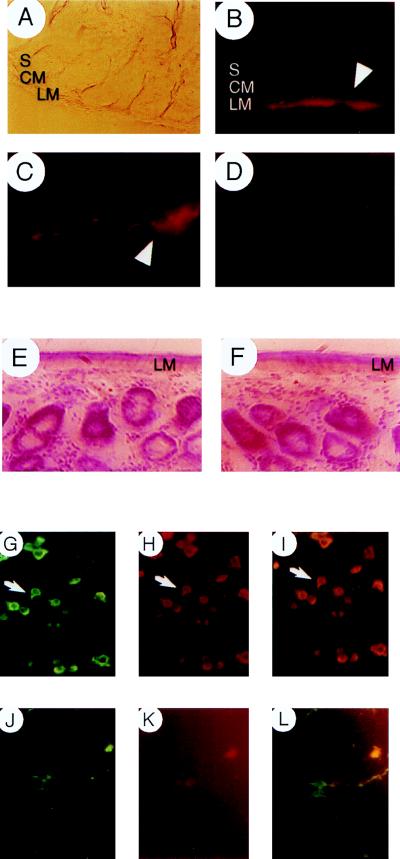
HO-2 and nNOS expression in the enteric nervous system. (A) Nomarski view of ileum from wild-type mouse. S, submucosa; CM, circular muscle; LM, longitudinal muscle. (B and C) HO-2 expression in myenteric ganglia (arrowheads) in wild-type mice. (B) Low power magnification. (C) High power magnification. (D) No HO-2 immunoreactivity is observed in HO-2 Δ/Δ mice. (E and F) Cross-sections of ilea from (E) wild-type and (F) HO-2 mutants stained with nuclear red for histochemical analysis. (Bottom) Colocalization of HO-2 and nNOS. Doubly labeled myenteric ganglia (arrows denote example) from colchicine-treated ilea of wild-type mice with (G) anti-HO-2 and (H) anti-nNOS, double exposure (I). Rat primary cortical neurons doubly labeled with (J) anti-HO-2 and (K) anti-nNOS, double exposure (L). For additional controls, see Materials and Methods. Double labeling with two different NOS antibodies yielded similar results.

