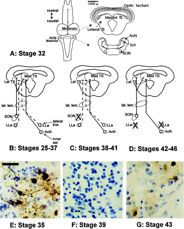
Figure 3.
(A) Schematic representation of major auditory nuclei in the tadpole brainstem as derived from sections from a stage 32 tadpole. Scale bar represents 1000 μm and refers to section outlines on right. Up until approximately stage 38, the tectal ventricle is continuous with the third ventricle. Connectivity pattern is based on retrograde transport of HRP after injections into the medial or lateral TS. Solid lines indicate auditory pathways; dashed lines indicate lateral line pathways. X, loss of retrograde cell filling in medullary brainstem nuclei that previously showed connectivity to the TS. NVIII, eighth (auditory) cranial nerve. (B–D) Changes in afferent connectivity across metamorphic development based on retrograde cell filling of afferents after HRP iontophoresis into the TS. The TS shows changes in overall morphology across metamorphic development, with the two nuclei of the medial torus not joining until stages 38–41. (B) Tadpoles from stage 25–37 (n = 13) showed robust filling of ipsilateral SON cells and cell filling in LLa bilaterally after injections in the lateral TS. HRP injection in the medial TS resulted in cell filling in the SON and in the contralateral AcN. (C) Tadpoles in the deaf period (n = 8) showed labeling of LLa neurons similar to that observed in earlier stage tadpoles but reduced labeling of the AcN and loss of labeling in the SON. (D) Tadpoles in metamorphic climax (n = 8) showed re-establishment of connectivity between the TS and the SON and increased labeling in the AcN but with a stage-dependent decrease in labeling of LLa neurons. By stage 44, no LLa neurons were labeled, and the AcN had expanded dorsally and medially into the region formerly occupied by the lateral line neuropil, coincident with the degeneration of the lateral line system at this stage. (E–G) HRP cell filling of ipsilateral SON neurons derived from injections into TS sites. Data are derived from injections matched for injection site, concentration of HRP, electrode diameter, and survival time. (Scale bar = 40 μm.) (E) Stage 35 tadpole, showing robust labeling of small round cell bodies from the SON. (F) Stage 39 tadpole showing lack of labeling of SON cells. (G) Stage 43 tadpole showing filling of SON cells demonstrating re-establishment of SON–TS afferent connectivity.

