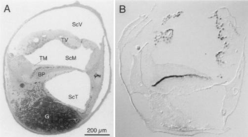
Figure 1.
Expression of the α1D subunit in cochlear hair cells. (A) Structure of the chicken’s cochlea. A semi-thin transverse section was taken near the middle of the organ and stained with toluidine blue. The hair cells are the dark, oblong cells at the upper margin of the basilar papilla (BP), the homolog of the mammalian organ of Corti; tall hair cells are toward the left and short hair cells are toward the right. ScV, scala vestibuli; ScM, scala media; ScT, scala tympani; TV, tegmentum vasculosum; TM, tectorial membrane; G, cochlear ganglion. (B) Localization of α1D mRNA by in situ hybridization. Digoxigenin-labeled antisense RNA derived from clone pBr64B was hybridized to a cryosection from a position similar to that in A and detected with anti-digoxigenin antibodies in a color reaction.

