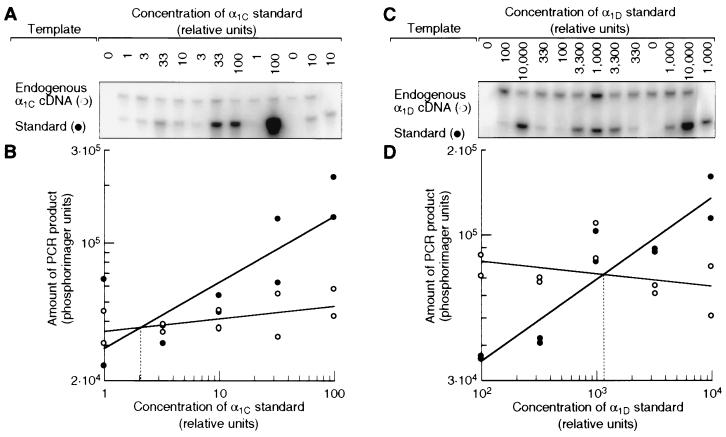
Figure 4.
Relative abundances of α1C and α1D mRNAs in the basilar papilla. (A) Southern blot of quantitative PCR assay. Aliquots of cDNA from the basilar papilla were spiked with serial dilutions of an α1C template as an internal standard. The PCR product of this standard was shorter than that of the endogenous α1C cDNA but accumulated with the same efficiency (data not shown). Both templates were amplified together with a primer pair specific for the α1C subunit, and the products were detected by hybridization with a radiolabeled oligonucleotide. The result from one of two independent experiments is shown. The control PCRs for the first and last lanes contained no cDNA from the basilar papilla. (B) Amounts of PCR products from endogenous α1C cDNA (○) and from α1C standard (•) plotted against the initial standard concentration. Straight lines fitted to each data set intersect at the standard concentration that gave rise to the same amount of PCR product as the endogenous α1C cDNA (dotted line). This standard concentration therefore was equal to the concentration of endogenous α1C cDNA in the sample. (C and D) Same as A and B, but with primers and standards for the α1D cDNA. The relative unit of concentration represents the same number of template molecules for both standards.

