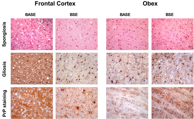
Figure 2. Histopathology and PrPres immunostaining.
Spongiosis, gliosis (GFAP staining) and PrPres deposition in frontal cortex and obex in BASE- and cBSE-infected primates (original magnification ×200 for spongiosis and gliosis, ×400 for PrPres staining). Immunostaining of PrPres was performed with 3F4 monoclonal anti-PrP antibody after proteinase K treatment as previously described [11]. No staining was observed in the brain of control healthy primates (data not shown) in these conditions.

