Fig. 3.
Photomicrographs of cochlea from subject V:3.
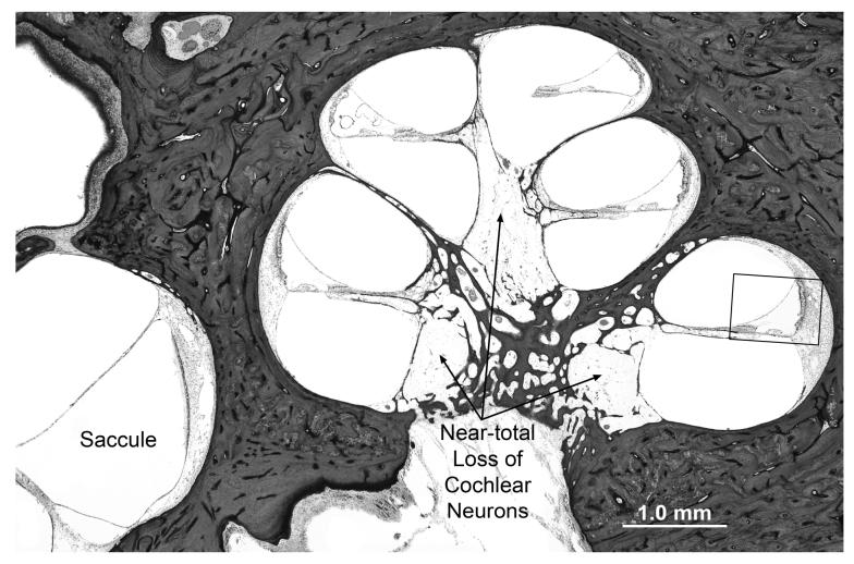
A. Lower power view showing cochlear turns and saccule. There is near total loss of spiral ganglion cells with severe atrophy of both peripheral and central axonal fibers. The saccule appears normal. The boxed area of the basal turn is shown at higher magnification in B.
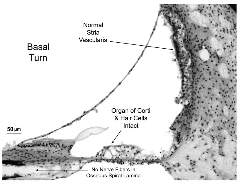
B. Higher power view of scala media from basal turn showing that the organ of Corti (including hair cells) is intact. Other structures of the cochlear duct such as stria vascularis, spiral limbus and tectorial membrane appear normal. Note the absence of nerve fibers in osseous spiral lamina.
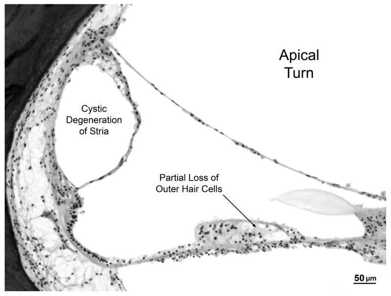
C. High power view of apical turn showing cystic degeneration of the stria vascularis and partial loss of outer hair cells within the organ of Corti.


