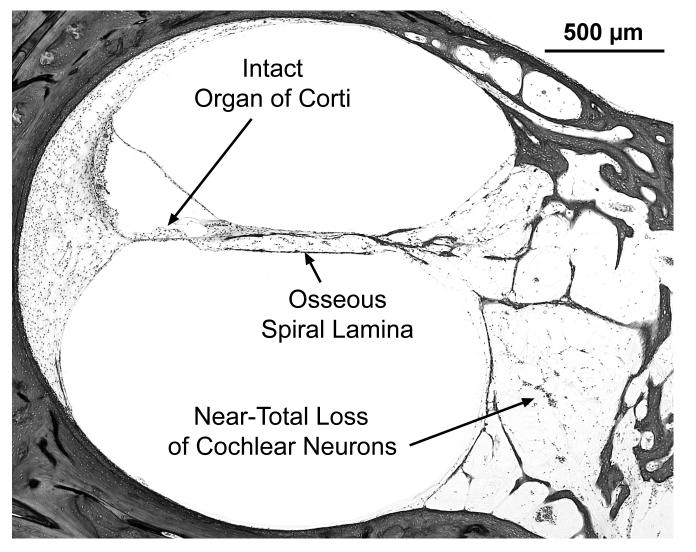
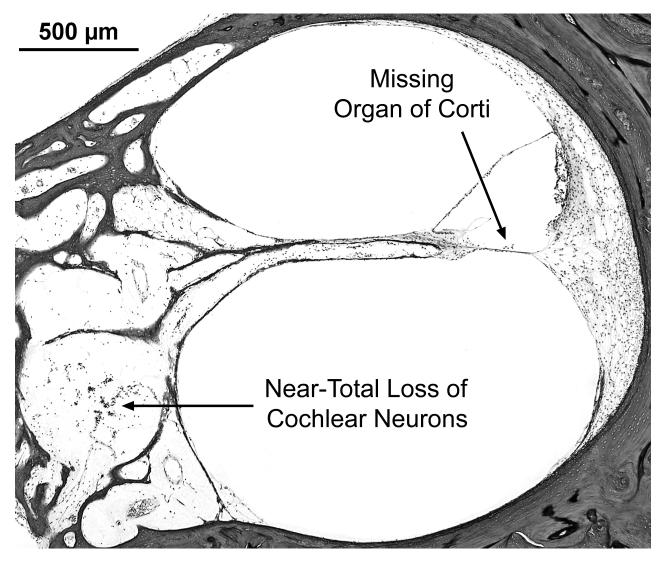
Fig. 4.
Subject VI:24. Views of scala media from basal turn in right (A) and left (B) ears of this subject. On the left, the organ of Corti is missing, whereas, it is intact and normal on the right. Both sides show near-total loss of cochlear neurons and loss of nerve fibers in the osseous spiral lamina.


