
Figure 2.
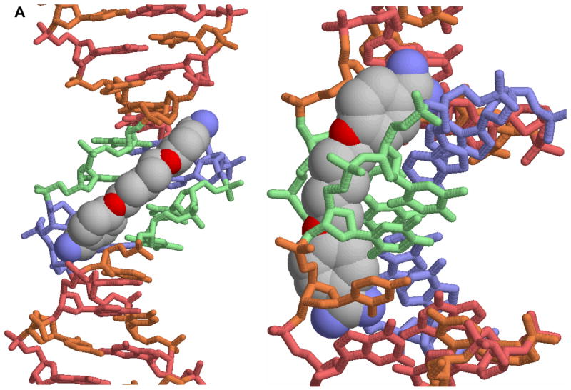

A. A view of the crystal structure of pentamidine bound to the -AATT- site of d(CGCGAATTCGCG)2 is shown [25]. The view on the left is into the minor groove and the view on the right shows how the compound fits into the groove. In this conformation the amidines are H-bonded to TO2 groups and the compound makes contacts with the groove floor and walls. B. A view into the minor groove of the DB75 crystal structure with the same binding site and DNA sequence is shown at the left [53]. The DNA contacts are similar to those with pentamidine but the DB75 structure is preorganized to bind in this orientation. The view on the right is from the major groove through the base pairs to DB75. The red dots identify the amidine to TO2 H-bond interactions. The compound curvature allows DB75 to slide deeply into the groove in AT sequences and make excellent contacts with the bases and groove walls.