Figure 4.
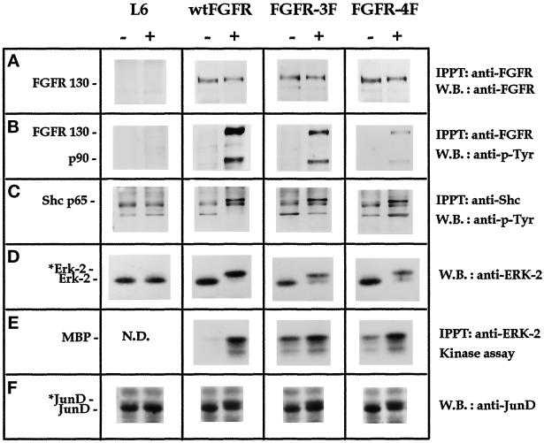
Signal transduction analysis of FGFR1 mutants. Parental L6 myoblasts, wt-FGFR1, FGFR1–3F, and FGFR1–4F cells were nonstimulated (−) or treated (+) either for 5 min (A, B, and C) or 20 min (D, E, and F) with 100 ng/ml FGF2. (A) Anti-FGFR1 immunoprecipitates were analyzed for FGFR1 expression using anti-FGFR1 antibodies. (B) Anti-FGFR1 immunoprecipitates were analyzed for tyrosine phosphorylation using antiphosphotyrosine (P-Tyr) antibodies. (C) Anti-Shc immunoprecipitates were analyzed for tyrosine phosphorylation. (D) Cell lysates (50 μg of total protein) were analyzed with anti-ERK2 antibodies. *Erk-2 indicates the phosphorylated protein. (E) Anti-ERK2 immunoprecipitates were analyzed for kinase activity using myelin basic protein (MBP) as substrate. (F) Cell lysates (50 μg of total protein) were analyzed with anti-JunD antibodies. *JunD indicates the phosphorylated protein. N.D., not determined.

