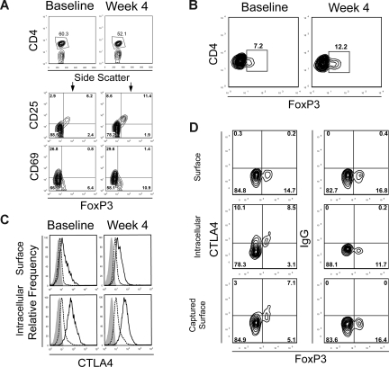
Figure 3.
Expansion of FoxP3+ CD4+ T cells after treatment. (A) PBMCs from baseline and at week 4 from a subject in dose level 5 were stained for CD4, CD25, and CD69 with fluorescence-labeled antibodies. Anti-FoxP3 antibody staining was performed after cell permeabilization. Cells were then analyzed by flow cytometry and gated on CD4+ T cells. CD4+ T cells were gated (top panels) and analyzed for CD25, CD69, and FoxP3 expression. The numbers on plots represent the percentage of cells in each of the quadrants. Data are derived from 1 subject treated in dose level 5 and are representative of 6 subjects in this cohort. Gating for CD4+ FoxP3+ staining was set with results from isotype-matched control IgG staining. (B) PBMCs from baseline and at week 4 from a subject who received anti-CTLA4 antibody at 3 mg/kg alone in a separate clinical trial for metastatic hormone-refractory prostate cancer were also stained for CD4 and intracellular FoxP3. Results are representative of 3 patients assessed. (C) PBMCs from baseline and at week 4 from a subject in dose level 5 were stained for CD4 and FoxP3 as described. Phycoerythrin-labeled anti-CTLA4 antibody, which is not blocked by the study drug, was also added either before (surface staining, top panels) or after (intracellular staining, bottom panels) cell permeabilization. FoxP3+ CD4+ T cells (solid line) or FoxP3− CD4+ effector T cells (dashed line) were gated and assessed for CTLA4 expression. Staining with an isotype-matched control antibody is also shown (shaded histogram). (D) Dynamics of the intracellular pool of CTLA4 were assessed by surface capture staining. PBMCs from a representative subject in cohort 5 at week 4 of treatment were cultured for 6 hours and stained with antibody for surface CTLA4 or intracellular CTLA4 (after permeabilization) at the conclusion of culture, or the cells were cultured in a cocktail containing monensin, brefeldin A, and an anti-CTLA4 antibody to capture surface CTLA4 that translocates to the cell membrane over this time. PBMCs were also stained for CD4 and FoxP3 with fluorescently labeled antibodies, assessed by flow cytometry, and gated on CD4+ T cells. Results are representative of PBMCs from 3 different patients in cohort 5 at week 4 or at baseline.

