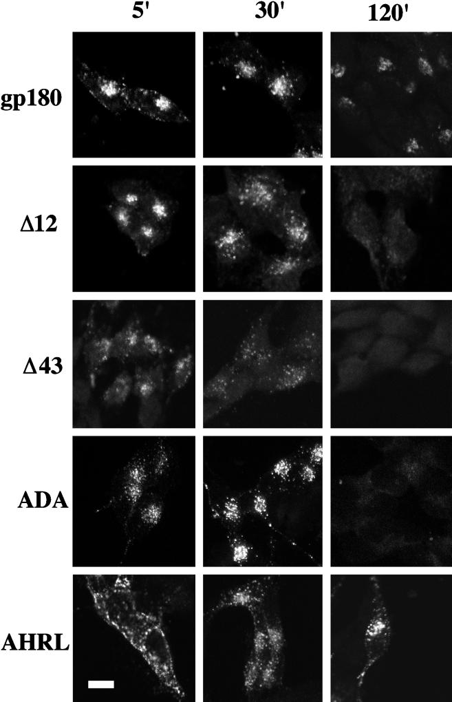
Figure 6.
Time-course analysis of antibody internalization. Stable transfectants expressing similar levels of gp180, the deletion constructs, or the point mutants were incubated with anti-gp180 antiserum at 4°C for 1 h, washed three times with cold PBS, and then incubated at 37°C for the times indicated in minutes (5′, 30′, and 120′). Cells were fixed, permeabilized, and stained with Texas Red-conjugated anti-rabbit IgGs from goat. Shown are the analyses of single clones. Bar, 10 μm.

