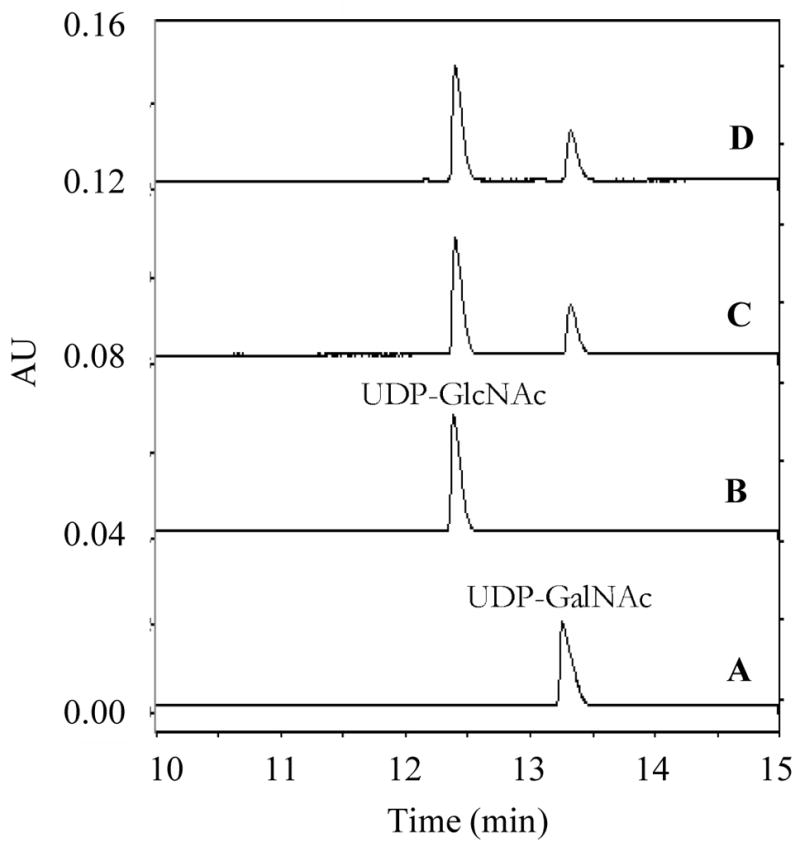
FIGURE 9.

Capillary electrophoresis separation of UDP-GlcNAc and UDP-GalNAc. The capillary was bare silica 75 × 57 cm with the detector set at 50 cm. The running buffer was 100 mM sodium tetraborate, pH 9.4. The separation was performed at 22 kV. Trace A, UDP-GalNAc (3) alone; Trace B, UDP-GlcNAc (2) alone; Trace C, UDP-GalNAc+TviC; Trace D, UDP-GlcNAc+TviC. The retention time of UDP-GalNAc (3) (trace A) and UDP-GlcNAc (2) (trace B) were 13.3 and 12.4 min, respectively.
