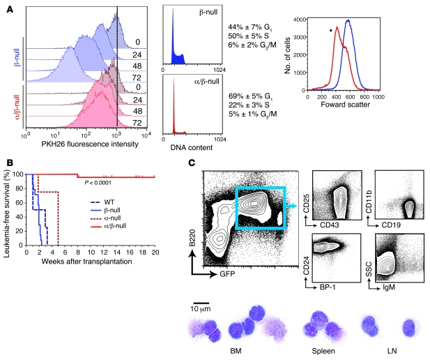
Figure 4. Severe loss of leukemogenic potential of α/β-null L-CFCs.
(A) Proliferation and cell cycle in α/β-null L-CFCs were assessed in vitro, where β-null (blue) and α/β-null (red) L-CFCs were labeled with 2 μM PKH26 and subsequently analyzed for dilution of PKH26 every 24 hours (left panel, representative of 2 independent experiments using 2 clones) and cellular size analyzed (right panel; *P < 0.05, 2-tailed, paired Student’s t test) by forward scatter; n = 3 clones. (B) Survival analysis of sublethally irradiated NOD/SCID mice transplanted with p190 L-CFCs (1 × 10⁶ cells). EGFP+ (or human CD4+) cells (>98% purity) from BM and spleen were collected to assess class IA PI3K deletion status (immunoblot analysis and leukemia-free survival were adjusted as described in Methods and Supplemental Figure 3 and Supplemental Table 1) (C) Representative immunophenotype and cytospins of β-null leukemic blasts in BM, spleen, and lymph nodes.

