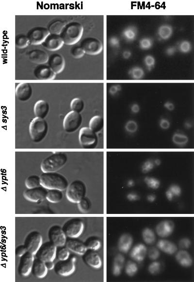
Figure 8.
FM4-64 staining of vacuolar membranes in ypt6/sys3 null mutants. Wild-type, Δsys3, Δypt6, and Δypt6/sys3 cells were grown at 25°C, labeled for 1 h with 30 μM FM4-64, washed three times with cold PBS buffer, and chased in YPD for 2 h. Then cells were examined by Nomarski and fluorescence microscopy for FM4-64 fluorescence. Essentially the same result was obtained after a 30-min chase time.

