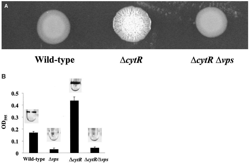
Figure 2.
Comparison of wild-type V. cholerae and ΔcytR mutant colony morphology and biofilm development.
A. Colony morphology of wild-type V. cholerae (MO10), a ΔcytR mutant (PW324) and a ΔcytRΔvps double mutant (PW329).
B. Biofilm accumulation by wild-type V. cholerae (MO10), a Δvps mutant (PW328), a ΔcytR mutant (PW324) and a ΔcytRΔvps double mutant (pw329). Biofilms stained with crystal violet are shown above, and quantification of crystal violet staining is shown below.

